Anatomy of the Occipital Sinus
About the illustration
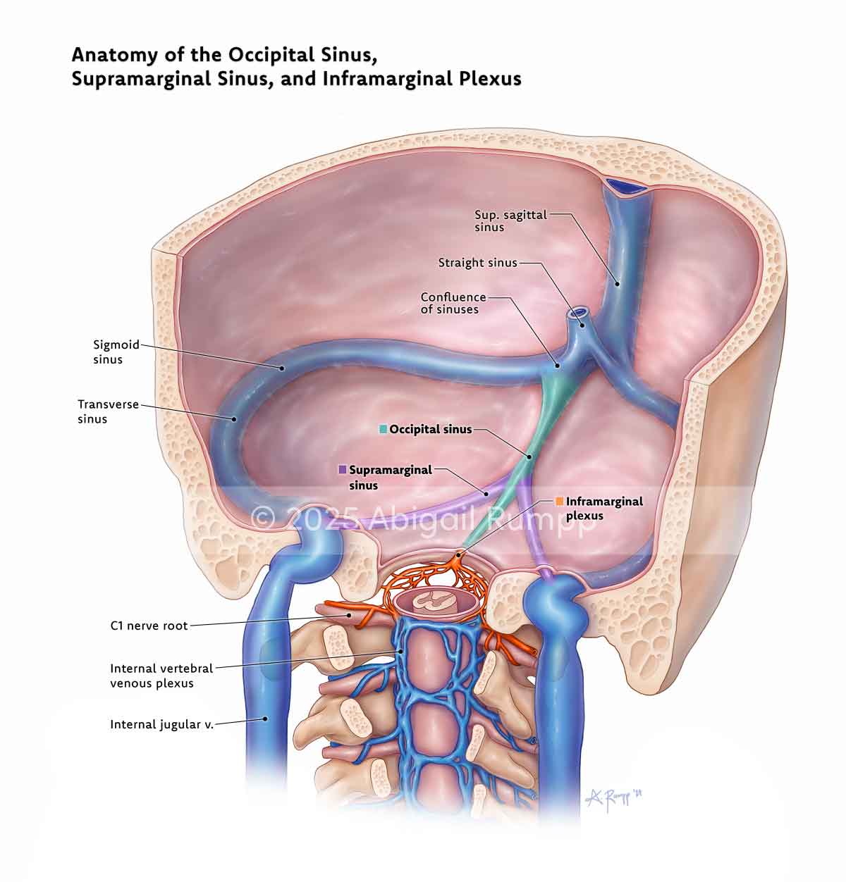
This illustration clarifies the venous anatomy surrounding the foramen magnum, with a focus on distinguishing between two distinct sinuses often conflated under the term “marginal sinus.” While commonly depicted as a single structure, the so-called marginal sinus is more accurately described as two separate components: the supramarginal sinus, which is intracranial, and the inframarginal sinus, part of the epidural spinal venous plexus located inferior to the foramen magnum and surrounding the C1 nerve roots. The illustration also depicts the occipital sinus—a small venous channel posterior to the confluence of sinuses—and its connections to both the supramarginal and inframarginal sinuses via the occipital marginal sinus and occipital vertebral sinus, respectively.
The illustration was created in collaboration with Dr. Phillipe Gailloud, and is intended as a teaching tool for interventional neuroradiologists to support clearer interpretation of venous structures at the craniocervical junction.
Photoshop, Horos, 3D Slicer
2024