Full Illustration

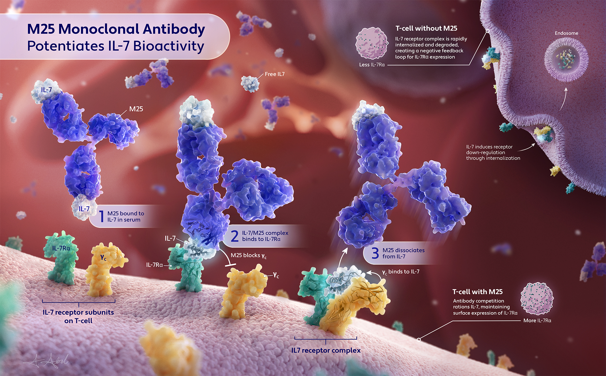
M25 Monoclonal Antibody Potentiates IL-7 Bioactivity

This illustration depicts the mechanism by which the M25 monoclonal antibody rations interleukin-7 (IL-7) to potentiate its cellular effects. IL-7 binds to the IL-7 receptor complex on T-cells. M25 rations IL-7 by blocking the binding of the common gamma chain, one of the proteins in the heterodimeric IL7 receptor complex. The 3 step IL-7 potentiation mechanism is illustrated on the foreground T-cell, highlighting this blocking. The inset in the upper left corner depicts typical IL-7 internalization in a T– cells not in the presence of M25. Smaller insets associated with each T-cell highlight the differences in IL7R-a expression between T-cell exposed to M25 and T-cells not exposed to M25.

CLIENT

Dr. Jamie Spangler, Emily Airail

ROLE

Scientific Illustrator: Responsible for visual concept development, layout design, 3D asset creation & rendering, and final illustration

AUDIENCE

Content experts

YEAR

2025

MEDIA

PyMol, ePMV Cinema 4D, Adobe Photoshop 

RESEARCH & THUMBNAILS

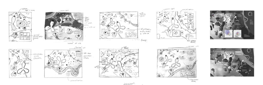
Thumbnailing helps organize information and provide visual previews for the client. Experimenting with different layouts and environments in the thumbnails helps me develop the most effective solution.  

Lorem ipsum dolor sit amet, consectetur adipiscing elit. Ut elit tellus, luctus nec ullamcorper mattis, pulvinar dapibus leo.

DRAFTS

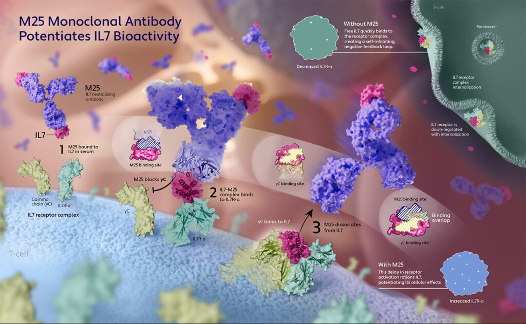
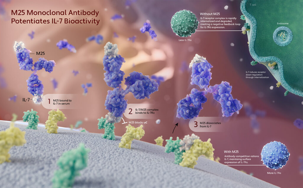
The vessel environment, general color scheme, and layout was solidified through the development of colored drafts based on the strongest thumbnails. Experimentation with insets, environmental color

Preliminary draft with binding site insets

Draft experimenting with T-cell color

LIGHTING & MATERIALS

Molecule materials were developed to match the color scheme of the molecules in the final submitted manuscript. As the environment developed, I adjusted the lighting to create a more realistic scenario, with the molecules appearing more backlit by the light from the end of the vessel.  

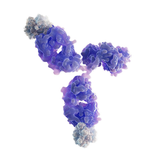
Antibody and IL-7 material test

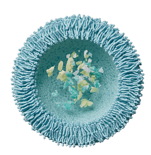
Endosome with degraded IL-7 and receptor complex

BEHIND THE SCENES

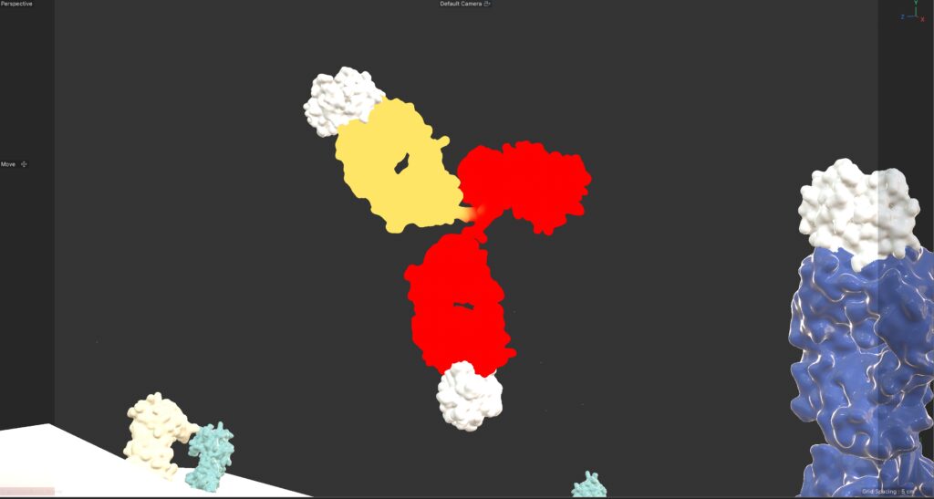
Because antibodies have flexible linkers connecting their Fc and Fab domains, I used vertex mapping and controllers to adjust these regions. This helped ensure the silhouette of the molecule remained identified in different binding configurations and created a more realistic scene. 

Antibody with vertex map