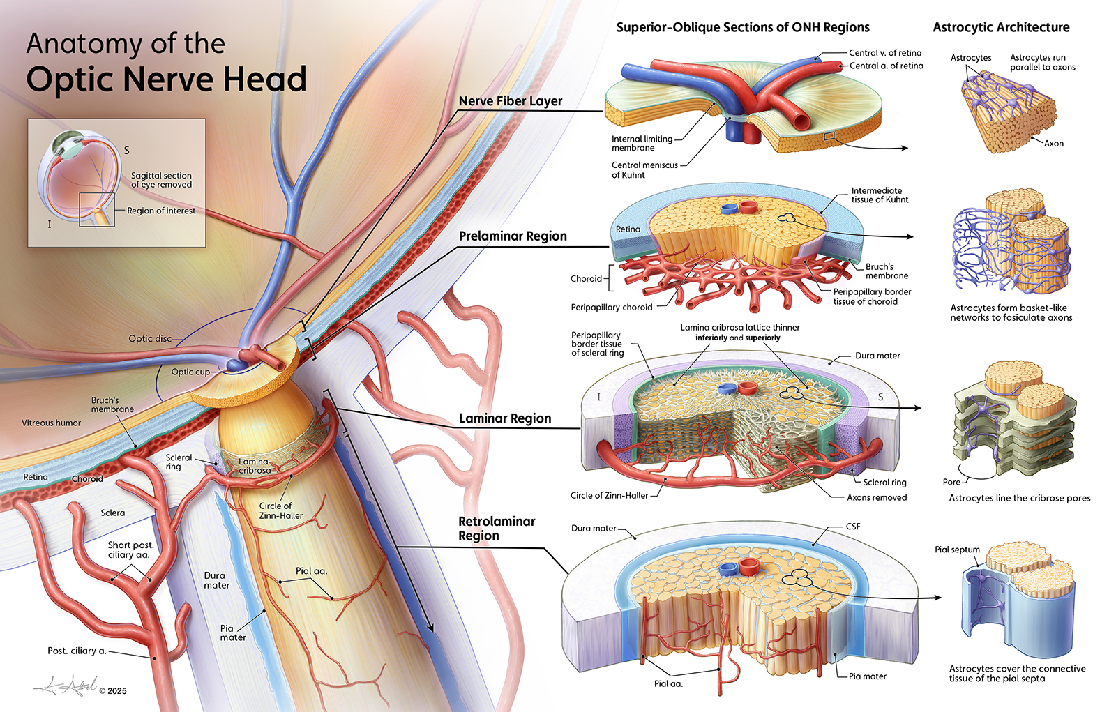
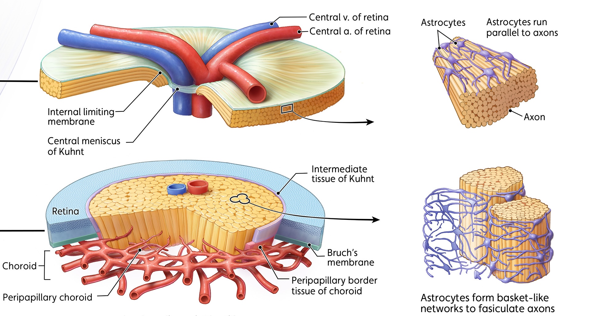
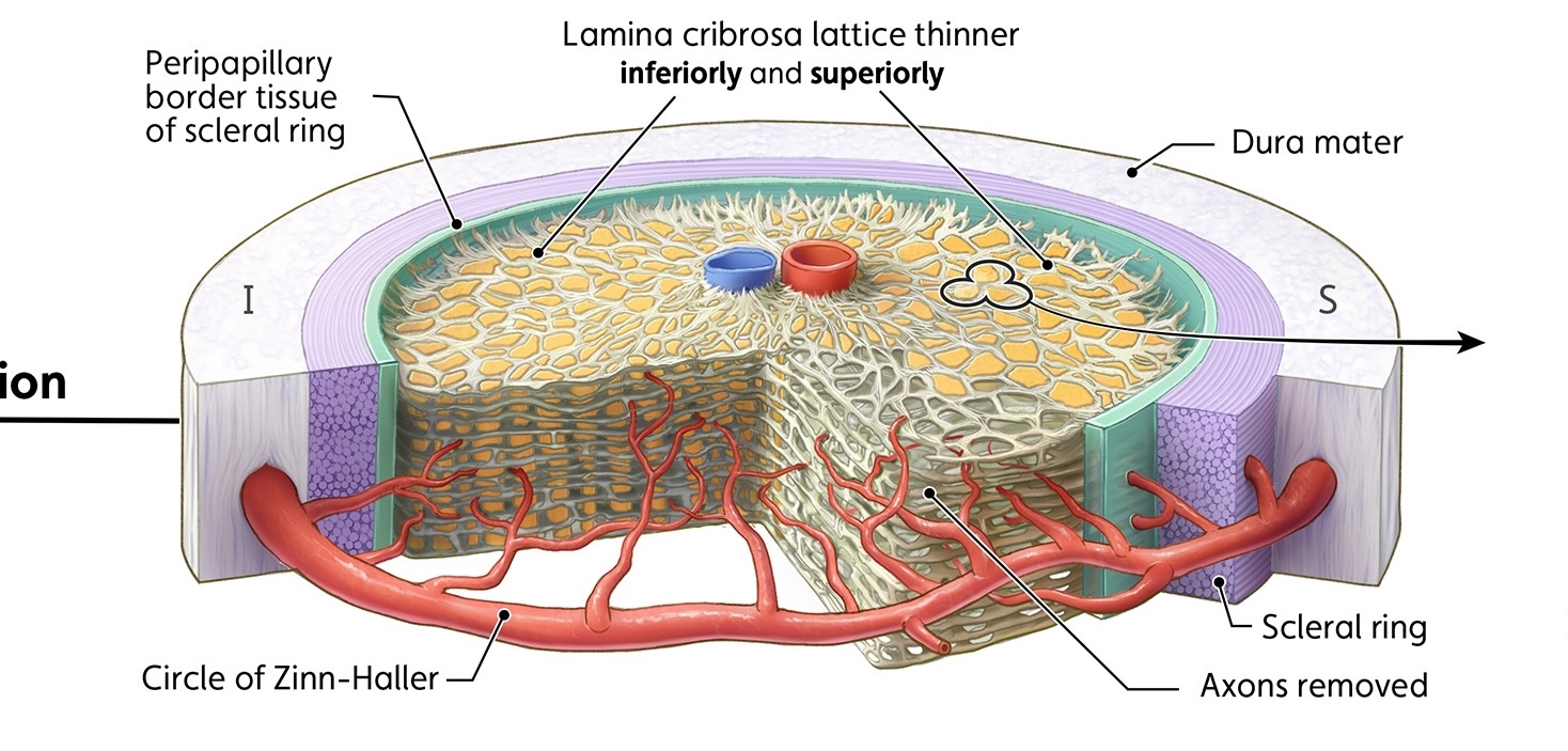
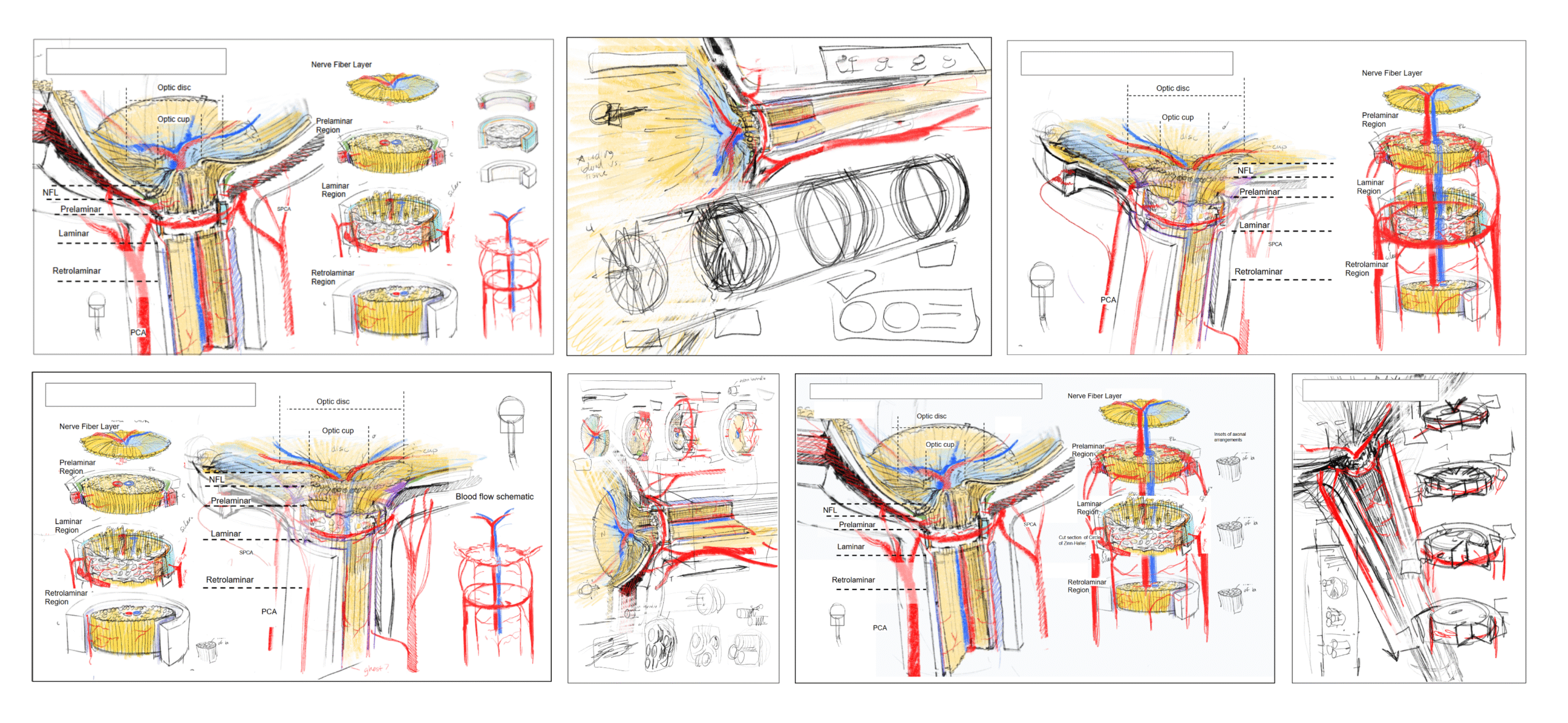
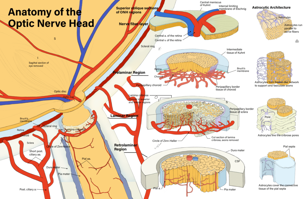
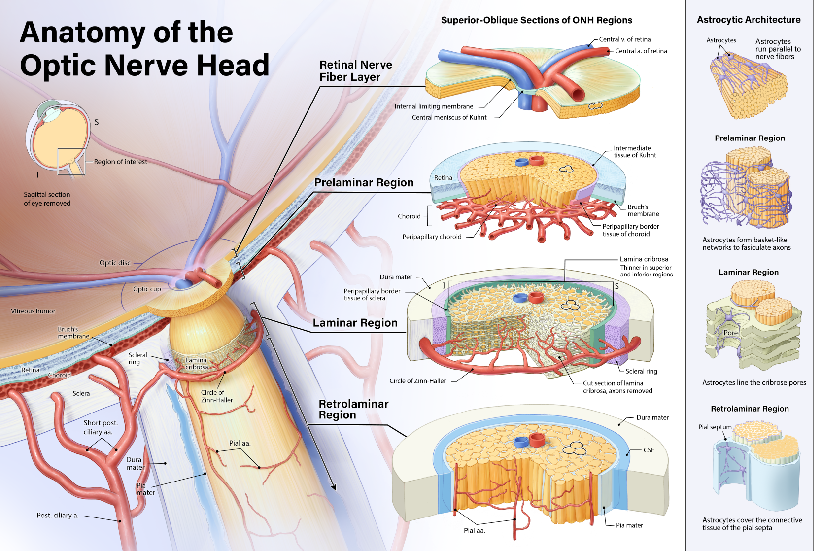
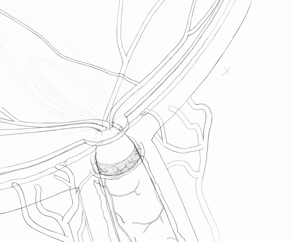
This anatomical illustration depicts the normal anatomy of the optic nerve head (ONH), highlighting each distinct region. A diagram in the upper left orients the viewer to the region of interest in the main image on the left, which depicts a sagittal section of the eye and optic nerve from a superior-oblique view. The optic nerve and a section of nerve fiber layer project outward from the section to illustrate their relative sizes in three dimensions. In the first column of insets to the right, a slice of each ONH region is isolated and enlarged to clarify the specific regional blood supply, unique structures, and the axonal organization characteristic of each region.
Each ONH region has a unique astrocytic organization that is important for regional classification. A section of 2-3 bundles of axons in each regional slice are featured in a second column of insets to illustrate this astrocytic organization.