Ultrasound-guided Fetal Embolization of Vein of Galen Malformation
YEAR
2025
CLIENT
Dr. Phillippe Gailloud, MD
Dr. Ahmet Baschat, MD
Johns Hopkins Medicine Division of Interventional Neuroradiology and Department of Gynecology and Obstetrics
MEDIA
Adobe Photoshop
DESCRIPTION
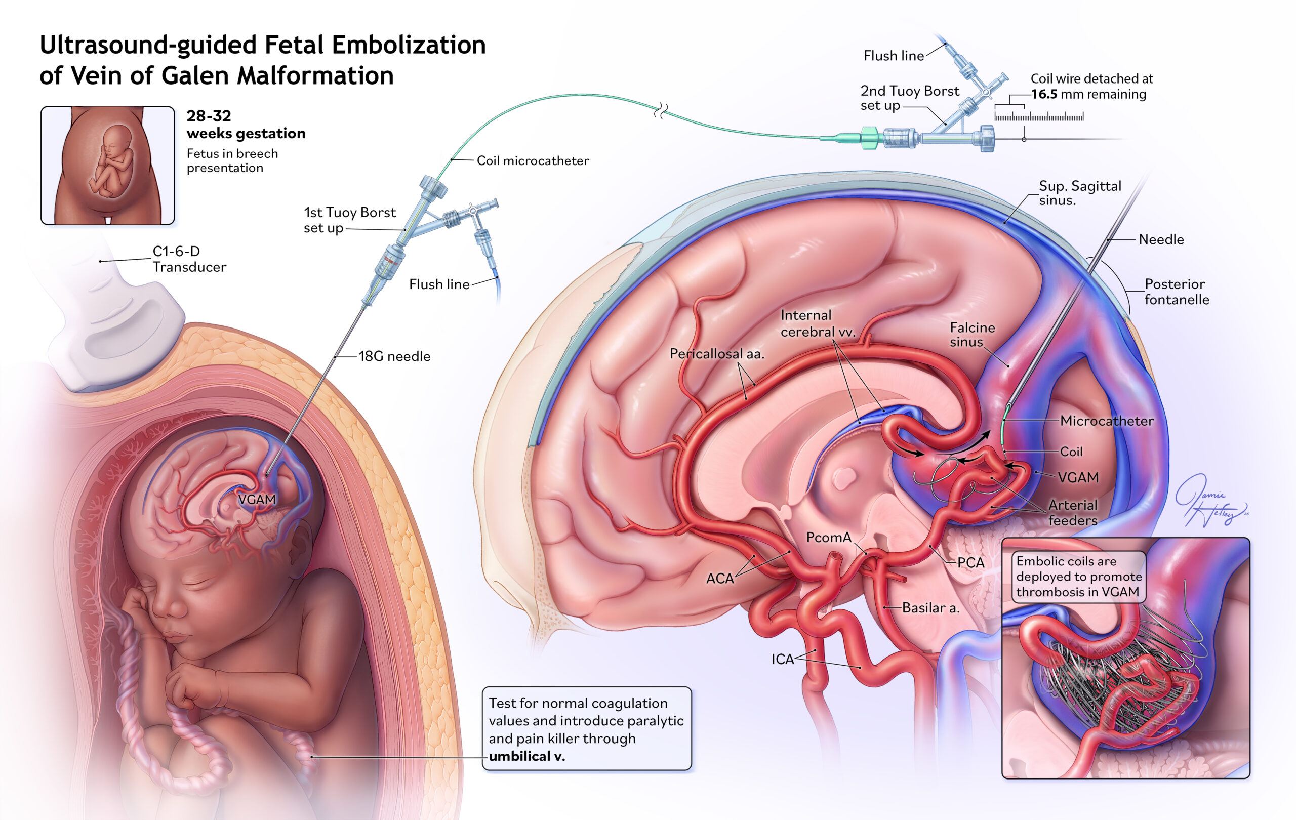
As one of the first cases of fetal cerebrovascular surgery, ultrasound-guided fetal embolization of a vein of Galen malformation marks a significant advancement in prenatal medicine, shifting management from postnatal intervention to prenatal treatment with the potential to reduce high neonatal mortality associated with this condition.
This illustration depicts the procedure and associated anatomy in detail: the upper left diagram introduces a fetus in breech presentation, while the section below shows the positioning of a curvilinear ultrasound transducer guiding a needle through the maternal abdomen, uterus, and into the posterior portion of the fetal head to reach the vein of Galen. The connected coil system is also shown. The main image places the malformation in context with the cerebrum and highlights the arteriovenous fistula, including enlarged feeding arteries such as the pericallosal and posterior cerebral arteries that directly connect to the vein of Galen. Approaching this illustration with a slightly oblique sagittal view provides clarity on the needle placement, microcatheter advancement, and coil delivery, while the right inset demonstrates how the density of deployed embolic coils slows blood flow and promotes thrombosis within the vein of Galen.