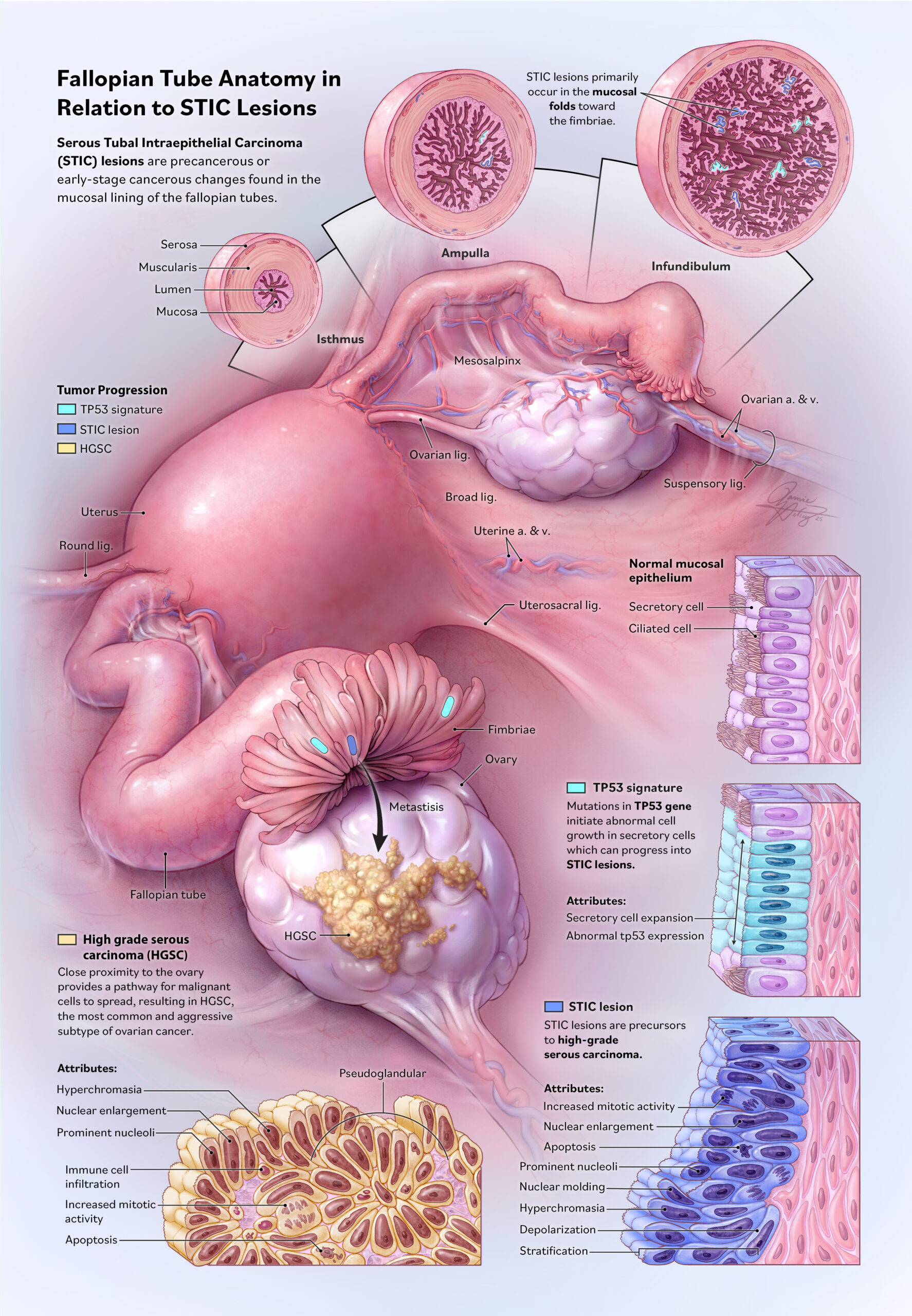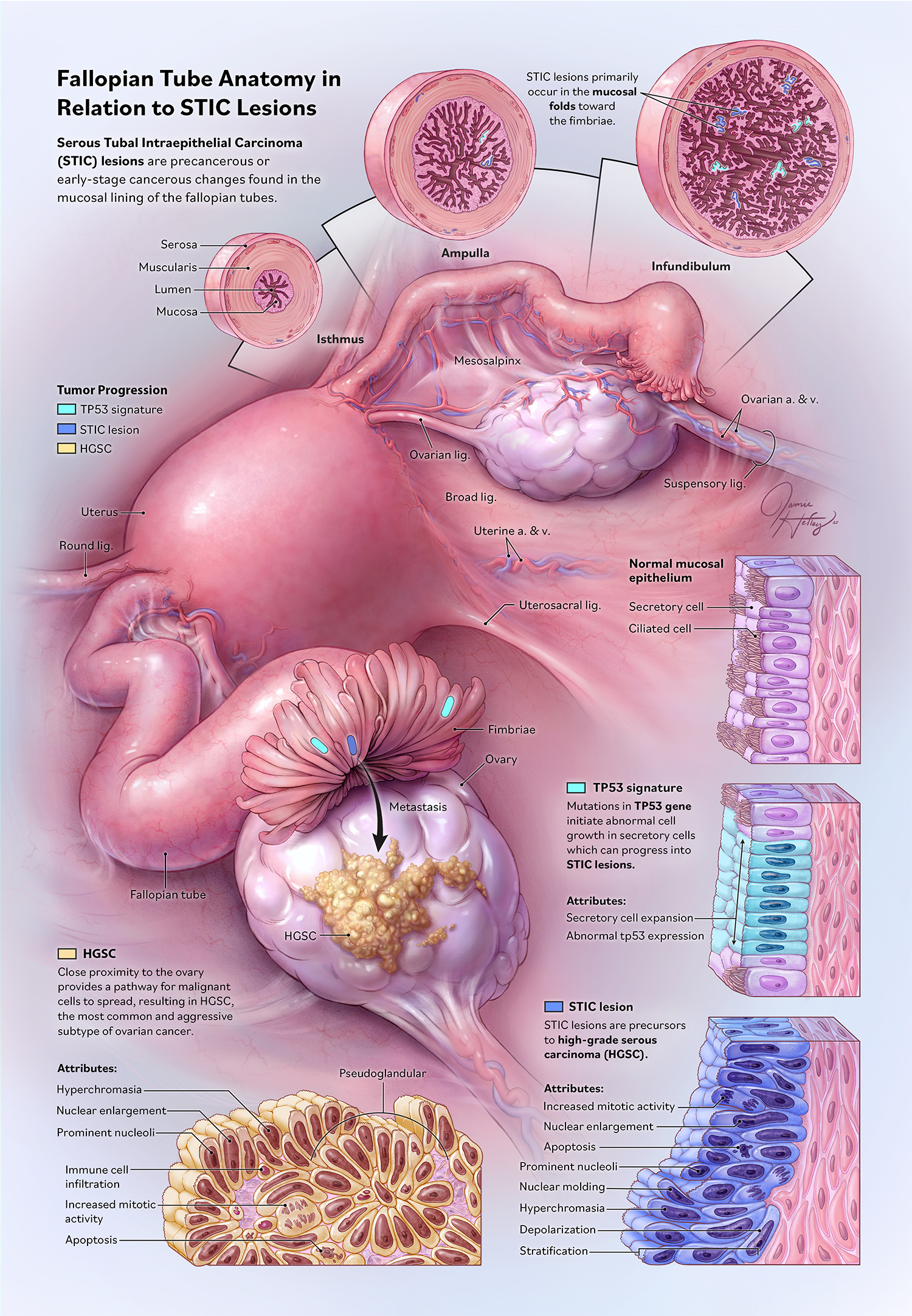
Fallopian Tube Anatomy in Relation to STIC Lesions
YEAR
2025
CLIENT
Dr. Brian Wildey
Johns Hopkins University
SOM Department of Gynecology and Obstetrics
Media
Photoshop
DESCRIPTION
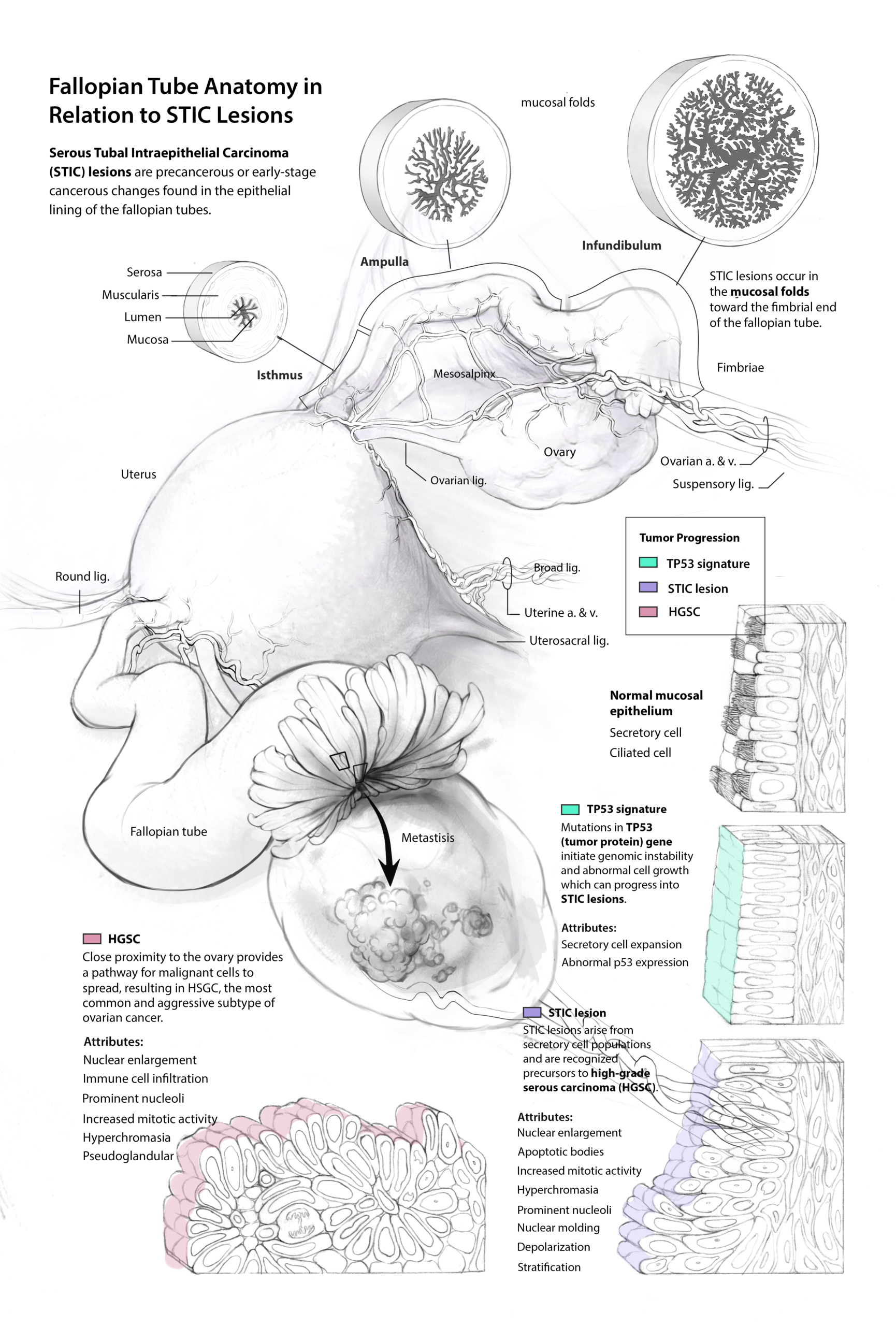
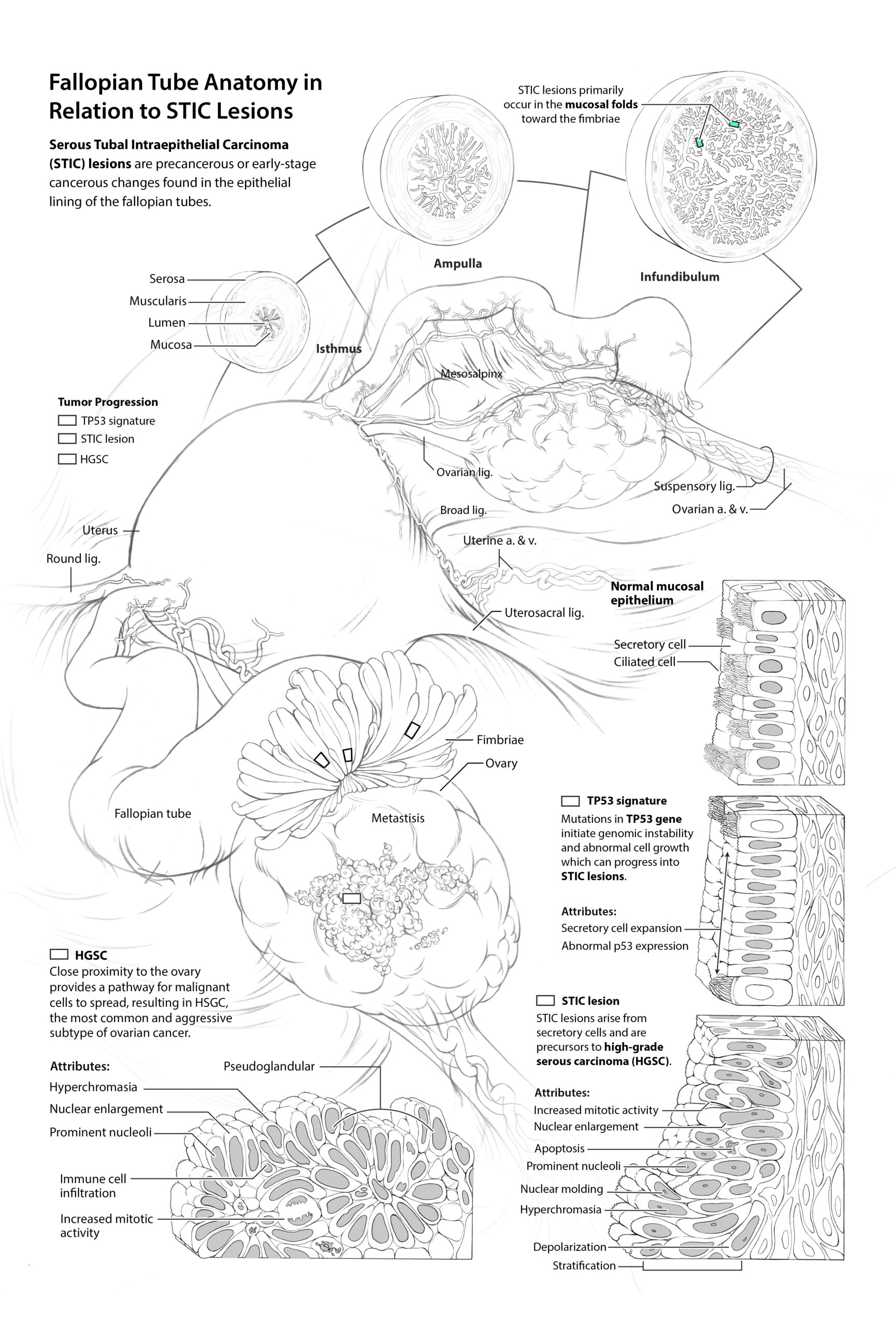
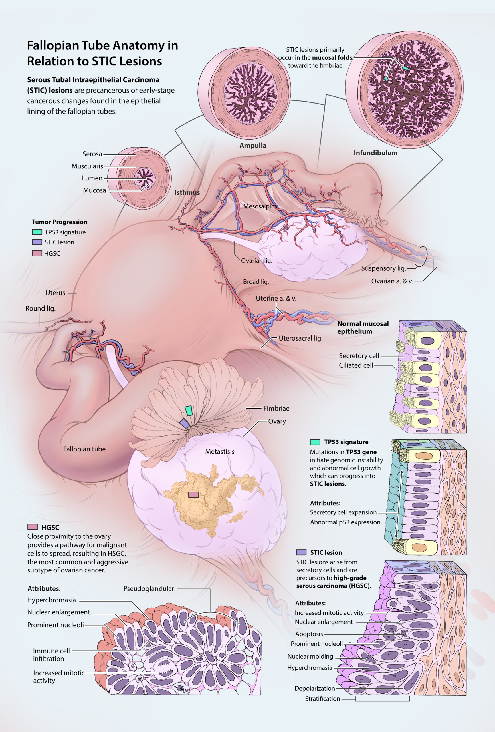
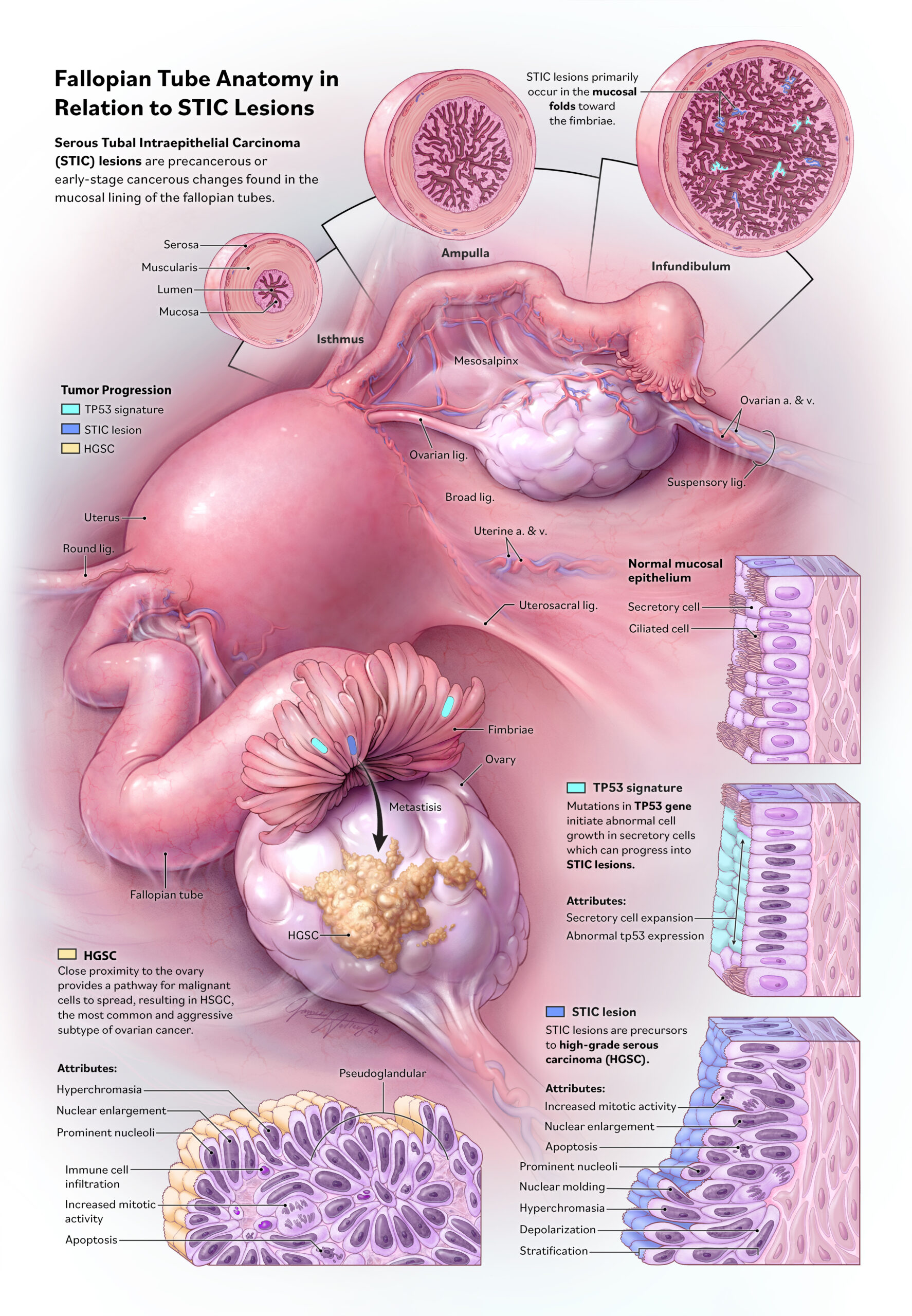
This illustration depicts the anatomy of the fallopian tube with the intent to enhance understanding of its relationship in the development of Serous Tubal Intraepithelial Carcinoma (STIC), a recognized precursor to Ovarian High-Grade Serous Carcinoma (HGSC).
The central illustration depicts the structure of the fallopian tubes and surrounding anatomy within the pelvic cavity, highlighting their spatial relationship to the ovaries. This visual representation aids in understanding how malignant transformations can originate in the fallopian tube epithelium and spread to adjacent structures. The insets above illustrate cross-sectional comparisons of the fallopian tube’s isthmus, ampulla, and infundibulum. These sections highlight the progressive complexity of internal mucosal folds toward the fimbriae where mutations in TP53 gene and STIC lesions primarily occur. The cellular diagrams positioned to the right and bottom follow the pathogenesis of HGSC. Didactic colors were used to highlight the morphological transitions from normal mucosal epithelium, TP53 signature, and precursor STIC lesions to HGSC. By clarifying these early changes, the illustration aims to improve understanding, support early detection, and inform preventative strategies for high-risk individuals.